


Download a PDF of the Handout by clicking on the image above.
 The Molecular Epigenetics Group (MEG) of the Life Sciences Institute at the University of British Columbia consists of 8 researchers from 3 departments (Biochemistry, Medical Genetics, and Zoology) with a common interest in epigenetic gene regulation.

X: A mosaic with XIST
Carolyn Brown’s laboratory studies the mechanism of human X chromosome inactivation – the process that equalizes the expression of X-linked genes between males and females.
X chromosome inactivation occurs early during mammalian development to transcriptionally silence one of the pair of X chromosomes in females, thereby achieving dosage equivalence with males who have a single X chromosome and the sex-determining Y chromosome. Research in the lab is directed towards understanding both the mechanisms involved in the inactivation process and the clinical implications of X chromosome inactivation in females.

Dr. Matthew Lorincz
Dr. Lorincz’s laboratory is focused on the interplay between transcription, DNA methylation and histone modification in development, using the mouse as a model system.
A number of factors have been described that catalyze the post-translational addition or removal of specific moieties, such as acetyl or methyl groups, to/from specific residues on the core nucleosomal histones, features of the so-called “histone code”. Methylation of lysine 4 of the H3 tail, which is associated with the promoter regions of both actively transcribed and “poised” genes, was recently shown to inhibit de novo DNA methylation, thus serving to “protect” promoter regions from DNA methylation. Conversely, we have shown that trimethylation of H3K9 (H3K9me3), a mark associated with transcriptional repression that is inversely correlated with H3K4me3, is enriched in embryonic stem cells at the promoter regions of a subset of germline-specific genes and specific endogenous retroviruses ERVs. This mark is deposited by the H3K9 KMTase Setdb1, generally independent of DNA methylation (See Matsui et al, Nature, 2010) and serves to maintain a number of ERVs in a silent state in cells deficient in all three DNA methyltransferases (See Karime et al, Cell Stem Cell, 2011). Surprisingly, HP1 proteins, “readers” of the H3K9me3 mark, are dispensable for H3K9me3-mediated proviral silencing (see Maksakova et al, Epigenetics & Chromatin, 2011) raising the question, how does H3K9 methylation inhibit transcription?
Ongoing research in the lab is directed towards characterizing the interplay between readers and writers of covalent histone marks, chromatin remodeling factors and DNA methylation in transcriptional regulation of genes and retroelements (see Thompson et al, PLoS Genetics, 2015 and Sharif et al. Cell Stem Cell, 2016), using knock-down, conventional and CRISPR-based genetic knock-out approaches. Employing Illumina next generation sequencing and bioinformatics pipelines developed in house (see Younesy et al, Bioinformatics, 2014), we systematically characterizing the role of histone H3K9 methyltransferases, H3K9me “readers” and chromatin remodeling factors in ESCs via RNAseq, ChIPseq, meDIPseq and hmeDIPseq analyses (see Liu et al., Genes & Development, 2014).
Exploiting genome-wide analyses and CRISPR-based deletion of specific LTR elements in the mouse genome, we are also characterizing the function of LTR elements as alternative genic promoters in ESCs and early embryos as well as germ cells (see Thompson et al. Molecular Cell, , 2016) and studying the impact of specific elements on the transcriptome in distantly related mouse strains as well as in rats.
We also recently developed an ultra-low-input micrococcal nuclease-based native ChIP (ULI-NChIP) and sequencing method (See Brind’Amour et al, Nature Communications, 2015) to generate genome-wide histone mark profiles with high resolution from as few as 103 cells. We demonstrate that ULI-NChIP-seq generates high-quality maps of covalent histone marks from 103 to 106 embryonic stem cells. Subsequently, we showed that ULI-NChIP-seq H3K27me3 profiles generated from E13.5 primordial germ cells isolated from single male and female embryos show high similarity to recent data sets generated using 50–180x more material. We are currently applying this method to address fundamental questions about intergenerational inheritance of covalent histone marks and their role in enhancer function and the inheritance of DNA methylation in the early mouse embryo.
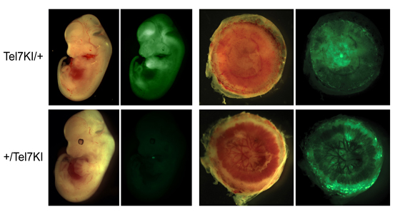
The research group of Louis Lefebvre studies the phenomenon of genomic imprinting, an epigenetic system guiding the monoallelic, parent-of-origin dependent expression of specific genes in mammals.
In placental mammals, both maternal and paternal genomes are required for normal development. This non-equivalence of the parental genomes is thought to represent the main barrier against parthenogenesis in mammals. This form of asexual reproduction in which only maternal DNA contributes to the offspring is frequently observed in insects and reptiles. In mammals, the parental genomes acquired at fertilization carry different epigenetic marks which are required for normal development. Genomic imprinting refers to this differential epigenetic marking of mammalian chromosomes in the male and female germ lines. One of the consequences of these differences between paternal and maternal homologues is the mono-allelic expression of certain genes according to their parent of origin. Unlike most genes used during development, imprinted genes are therefore only expressed from one allele, the other one being silenced by epigenetic marks such as DNA methylation and histone modifications. Because of imprinting, mutations in imprinted genes can therefore behave as dominant mutations, if inherited from the expressed allele, or as recessive mutations, when present on the silent homologue. A leading evolutionary theory to explain the emergence of imprinting in mammals proposes that imprinted genes regulate the exchange of nutrients between the pregnant female and her offspring during in utero development and the early postnatal period. The finding that imprinting seems to have evolved in lineages which exhibit placentation and that several imprinted genes are implicated in the regulation of embryonic growth and placentation all support this model.
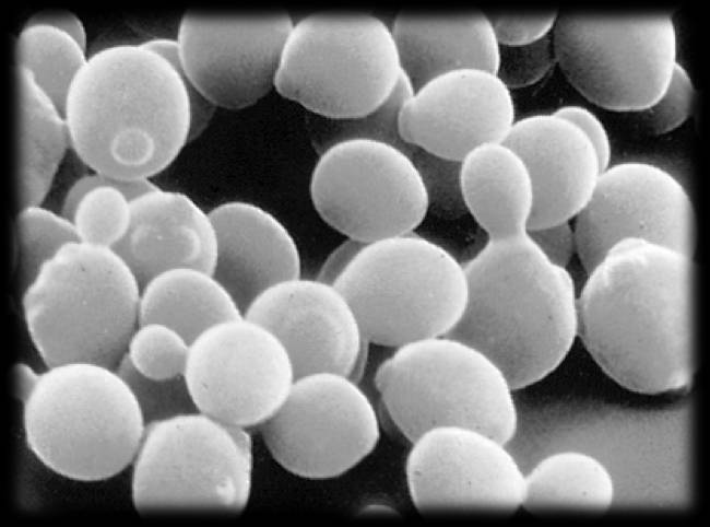
This research group studies the recruitment of histone modifying complexes to transcriptionally active genes in yeast.
Chromatin is a nucleoprotein structure, consisting of DNA, histones, and non-histone proteins, which packages DNA in the eukaryotic nucleus. Our research focuses on multi-protein complexes which post-translationally modify histones. We are interested in determining how these complexes are targeted to specific regions of the genome, and the functional consequences of this targeting. We use the budding yeast, Saccharomyces cerevisiae , as a model system due to its well-defined genetic system and the fact that there are a large number of eukaryotic genes that have been evolutionarily conserved between yeast and mammals.

Cancer cells and cells in developing embryos share two important characteristics: they rapidly proliferate and they are capable of migrating extensively. Because of this, it has been suggested that perhaps cancers are formed when adult cells mistakenly trigger dormant embryonic programs. In our lab, we are addressing this hypothesis using pigment cells, called melanocytes. Using classical genetic techniques, we seek to identify new genes that play a role in melanocyte development, to better understand the basic processes of cell migration, differentiation, survival and proliferation. We then study the role of these genes in human melanomas, cancers of melanocytes, which are increasingly common in Canada.

This laboratory studies how environmental signals affect transcriptional regulation in yeast using a combination of biochemical, molecular and genetic analysis.
Normal cell growth and development are highly regulated by hormones and factors from the environment, and cells respond to these signals by regulating expression of appropriate genes. Many diseases are caused or influenced by alterations in the relationship between cell signaling and gene regulation. For example, development of cancer can largely be attributed to uncoupling of gene regulation from growth factor signaling. Similarly, the pathology of AIDS is strongly influenced by control of HIV-1 gene expression through T cell activation signals. To elucidate fundamental mechanisms controlling signal responsive expression, we study the regulation of specific transcription factors in human T cells and the model eukaryotic organism Saccharomyces cerevisiae . Our research has been instrumental in revealing how responses to multiple signals can be coordinated through the function of the RNA polymerase II-associated protein kinase CDK8. Additionally, we have identified a human transcription factor that regulates establishment of latent HIV-1 provirus and controls reactivation of viral replication in response to T cell signaling.